

Gamma oscillation is the synchronization with a frequency of 30–90 Hz of neural oscillations, which are rhythmic electric processes of neuron groups in the brain. The inhibitory interneuron network is necessary for the production of gamma oscillations, but certain disruptions such as brain inflammation, oxidative stress, and metabolic imbalances can cause this network to malfunction. Gamma oscillations specifically control the connectivity between different brain regions, which is crucial for perception, movement, memory, and emotion. Studies have linked abnormal gamma oscillations to conditions of the central nervous system, including Alzheimer’s disease, Parkinson’s disease, and schizophrenia. Evidence suggests that gamma entrainment using sensory stimuli (GENUS) provides significant neuroprotection. This review discusses the function of gamma oscillations in advanced brain activities from both a physiological and pathological standpoint, and it emphasizes gamma entrainment as a potential therapeutic approach for a range of neuropsychiatric diseases. It also focuses on the fact that the US power grid and its reliance on 60hz oscillation fields may be affecting how our nervous systems work.
The provided sources touch on various aspects of electromagnetic fields and their effects on the brain. The Legros et al. study provides empirical evidence that 60 Hz magnetic fields can modulate brain activity as measured by fMRI, even without impacting task performance. The “Pasted Text” source offers a basic overview of brain waves and links power grid frequencies to potential nervous system stimulation. The Ichim et al. review delves into the complexities of gamma oscillations, their potential therapeutic roles (GENUS), and the hypothesis (GAMER) that endogenous gamma rhythms are vital for maintaining brain health. The combined information suggests a need for further investigation into how ELF EMFs interact with brain activity and the potential implications for both neurological function and therapeutic interventions. It is worth noting that Ichim et al. raises some doubt about the role of 40Hz stim, but also alludes to the positive results seen from it. Finally, there is no hard evidence or broad medical consensus on how ELF fields and frequencies influence consciousness and awareness, which is only anectodal at this time.
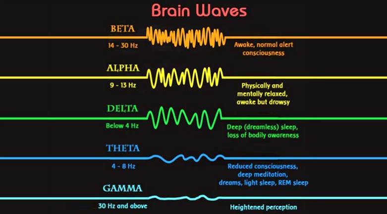
Brain waves are electrical impulses that flow through the brain, creating patterns of activity. They are measured in cycles per second, or hertz (Hz). There are four main types of brain waves: alpha, beta, delta, and theta.
Each type of brain wave is associated with a different state of consciousness. Beta waves, for example, are associated with wakefulness and alertness, while delta waves are associated with deep sleep.
Here’s a breakdown of the different types of brain waves and their associated frequencies:
Beta Waves (12-30 Hz)
– These brain waves are associated with alertness and wakefulness. They are most commonly observed in the frontal lobe of the brain.
Alpha Waves (8-12 Hz)
– These brain waves are associated with relaxation and calmness. They are most commonly observed in the occipital lobe of the brain.
Theta Waves (4-8 Hz)
– These brain waves are associated with meditation, creativity, and dreaming. They are most commonly observed in the temporal lobe of the brain.
Delta Waves (0.5-4 Hz)
– These brain waves are associated with deep sleep and unconsciousness. They are most commonly observed in the parietal and frontal lobes of the brain.
Gamma Waves 30-90Hz
Generation of gamma oscillations
Neural oscillations are rhythmic fluctuations generated by the activity of local neuron populations or neuron assemblies across brain areas and can be detected by local field potential (LFP), electrocorticography (ECoG), electroencephalography (EEG), and magnetoencephalography (MEG) at frequencies including delta (1-4 Hz), theta (4–8 Hz), alpha (8–12 Hz), beta (15–30 Hz), gamma (30–90 Hz) and high gamma(>50 Hz; Mathalon and Sohal, 2015; Cole and Voytek, 2017). They govern the timing of neuronal spikes at the microscale, and at the macroscale, they coordinate the dispersed cortical communication to enable temporal and spatial brain connectivity (Zhang et al., 2018). According to Pascal Fries’s hypothesis of “communication through coherence (CTC),” effective synaptic communication is dependent on the coordination between presynaptic and postsynaptic groups (Fries, 2015). Gamma oscillations, characterized as a rapid rhythm, allow excitation in the network to temporarily escape from the following inhibition, so enhancing the effectiveness, precision and selectivity of communication between multiple regions (Tiesinga et al., 2004). Electrophysiological data from macaques showed that virtually induced gamma synchronization between primary visual cortex (V1) and higher visual cortex (V4) facilitated sensory transmission to motor responses and shortened their reaction time, hence supporting the CTC hypothesis (Rohenkohl et al., 2018).
From The role of gamma oscillations in central nervous system diseases: Mechanism and treatment
Ao Guan 1,2,†, Shaoshuang Wang 1,†, Ailing Huang 3,†, Chenyue Qiu 3, Yansong Li 1, Xuying Li 1,3, Jinfei Wang 2, Qiang Wang 1,*, Bin Deng 1,3,*
PMCID: PMC9374274 PMID: 35966207
It is possible to externally adjust the rate of brainwaves through the process of entrainment. According to Gruzelier4, prolonged audio stimuli in repetitive and synchronized manner may induce changes in brain waves patterns and, consequently, may modulate neurophysiological and behavioral responses. More specifically, repetitive external or environmental stimuli may temporarily affect the predominance of specific brain wave frequencies, a phenomenon namely brainwave entrainment (BWE)5–7. Therefore, BWE can be defined as rhythmic synchronization of brainwave oscillation with an external repetitive stimulus.
BWE is a recurrent phenomenon in nature and biologically present in living beings8. The principle of entrainment or harmonization was discovered around 1665, by the Dutch scientist Christian Huygens9. The synchronization obtained through the entrainment principle is the result of the harmonization principle, a physical phenomenon that occurs systematically in nature, and that is dependent on environmental stimuli, for example, visual, auditory, or tactile. These stimuli may be used to elicit synchronized brainwave patterns to match that of different mental states and/or levels of consciousness, as seen with data acquisition techniques, such as the electroencephalography (EEG). In this context, Oster10 stated the possibility to improve, amplify or modulate brain wave patterns to conditioned events in the cerebral cortex3,11. The proposed therapeutic benefits have a wide scope, including the improvement of cerebral blood flow, neuroplasticity stimulation, and neurophysiological compensations between the cerebral hemispheres3.
It is also possible to control brainwave entrainment through harmonics of surrounding electrical fields. Clinical research indicates that somewhere between 25% and 75% of human and animal subjects may exhibit marked psychophysiological sensitivity to extremely low frequency (ELF) magnetic and electric fields.
Brain-wave entrainment can be demonstrated electroencephalographically when subjects are in the vicinity of oscillations in the frequency range of approximately 3–20 Hz at intensities below 100 nT (nanoteslas). (1 T = 104 G.)
ELF fields of 6.67 Hz, 6.26 Hz and lower tend to produce symptoms of confusion, anxiety, depression, tension, fear, mild nausea and headaches, cholinergia, arthritis-like aches, insomnia, extended reaction times, hemispheric EEC desynchronization, and many other vegetative disturbances. H and B field (magnetic vector) oscillations of 7.8, 8.0, and 9.0 Hz produce anxiety-relieving and stress-reducing effects that mimic some meditative states. It has been speculated that frequencies in this range may be the universally permeating
“clock frequencies” or carriers on which “mind” or “consciousness” states can be impressed and in which they may interact with other life-forms in the nebulous realms of ESP, psychotronics, distant healing, radiesthesia, and related paranormal but anecdotal phenomena.

Frequency of US Power grid
Short answer: The range is usually held within ±0.5%, so its from 59.7Hz to 60.3Hz for a 60Hz grid.
Long answer: Frequency is regulated tightly because it’s how the overall load in the grid is controlled. If there’s a runaway to lower frequencies, that usually means there is a short-circuit near a major power station or hub. So that will drop out soon. Then there usually is a runaway to higher frequencies because of the dropped load. So things escalate very quickly if the grid has many gigawatts power stations. And you don’t want that.
In weak grids on the contrary, the frequency may swing much more. Eastern European countries typically allowed 47Hz to 53Hz. That’s acceptable if there’s only a few power stations and a few big consumers. The same for emergency generators and isolated grids on islands.
What limits the frequency downwards is power conversion in transformers and AC motors. The lower the frequency is, the lower the primary voltage needs to be not to overexcite the iron parts by the magnetic fields. So a substantial frequency drop has to be accompanied by a voltage drop too, and that’s what is done in weak grids to allow lower frequencies.
Gamma waves above 50 are generated within very small regions of the brain associated with concentration of sensory awareness on external objects typically in visual centers, though strangely in olfactory centers for hedgehogs. Lower Gamma in the 20-40hz range may have a positive effect on concentration in alzheimer’s patients, but at what detriment to the neurologically necessary relaxation periods of lower brainwave frequencies. And specifically their interference in inducing states of meditation associated with lower frequencies like alpha and delta waves. The field of 60hz surrounds us with many people these days trying to protect themselves from EMF but how large is the em field and how far does it travel? Power lines differ in radiation emissions.
For street pole power lines with 33 kV, the strongest ones produce around 0.5 milligauss at a distance of 25 meters.
The lines with high voltage transmission lines of 400 kV create less than 0.5 milligauss at a 200-meter range.
The real estate industry would have you believe in these safety numbers What Is A Safe Distance to Live from Power Lines?
One of the most conflicting issues is measuring the safe distance from powerlines. It is still a highly controversial issue and depends on various facts. Another thing is the EMF effects depend on the distance of your house. So that after finding all the conflicting researches here is the safe distance guide-(About electromagnetic fields, 2020)(DC EMI EXPLAINED, 2020)
- For 133 kV Power lines you have to maintain 100 feet distance.
- For 230 kV Power lines you have to maintain 150 feet distance.
- For 345 kV Power lines you have to maintain 250 feet distance.
- For 550 kV Power lines you have to maintain 350 f feet distance.
After 500 feet it’s unable to measure the EMF effect with power lines. That is the EMF power lines safe distance. However, it’s still a question that will you feel risk-free to live in such an area?
But the entirety of the structures you are spending most of your time in are cycling the same HZ at 130 and 240 volts, so what is the safety distance from the wiring in your own home? This research is discouraged for obvious reasons lest we all go back to burning whale oil lamps at night. But despite all the technological miracle we’ve been living for the last 100 years with indoor wiring, it cannot help but have produced a change in our attention spans and externalized the focus of our attention to primarily visual stimuli.
Briefing Document: EMF Effects and Brain Activity
I. Electromagnetic Fields (EMF) and Measurement Considerations
- Source: InspectAPedia article on EMF measurement distance.
- Main Theme: This source focuses on practical considerations for measuring EMFs, particularly the importance of distance from the source.
II. 60 Hz Magnetic Fields and Brain Activation
- Source: “Effects of a 60 Hz Magnetic Field Exposure Up to 3000 μT on Human Brain Activation as Measured by Functional Magnetic Resonance Imaging – PMC” (Legros et al., 2015).
- Main Themes:fMRI Detects MF-Induced Changes: The study demonstrates the feasibility of using fMRI to detect subtle changes in brain activation patterns after exposure to 60 Hz magnetic fields.
- “These results illustrate the potential of using fMRI to identify MF-induced changes in functional brain activation, suggesting that a one-hour 60 Hz, 3000 μT MF exposure can modulate activity in specific brain regions after the end of the exposure period (i.e., residual effects).”
- Specific Brain Region Modulation: Exposure to a 60 Hz, 3000 μT magnetic field for one hour can modulate activity in specific brain regions.
- No Impact on Task Performance: Despite changes in brain activation, the study found no significant impact on the performance of finger-tapping or mental rotation tasks.
- “It should be noted that, despite the modulation in neuroprocessing presented in this paper, the 60 Hz MF exposure did not impact speed or accuracy of the tasks, and therefore did not have any physical behavioural impact.”
- Cortical Excitability: The study suggests that MF exposure may modulate cortical excitability, possibly via changes in synaptic plasticity.
- “We discuss the possibility that MF exposure at 60 Hz, 3000 μT may be capable of modulating cortical excitability via a modulation of synaptic plasticity processes.”
- Methodology:Participants performed finger-tapping and mental rotation tasks before and after a one-hour exposure to a 60 Hz, 3000 μT MF. Control groups were used.
- fMRI was used to measure brain activation patterns, focusing on Blood Oxygen Level Dependant (BOLD) signals.
- The magnetic field was generated using the MRI’s Z-gradient coil.
- Confounds:The study acknowledges that MRI scanners use various electromagnetic fields, which could potentially confound results. However, the consistent use of imaging sequences in both control and experimental groups helps to isolate the effects of the 60 Hz MF.
- The researchers took steps to measure and account for electric fields induced by the 60 Hz MF exposure in comparison to those induced by the fMRI BOLD sequence.
- Key Findings:Significant differences were found in task-dependent brain areas after MF exposure.
- Subjects could not consciously detect the presence of the 60 Hz MF.
- The study demonstrates that fMRI is a valuable tool for imaging the effects of ELF MF on human neurophysiology.
- It’s important to restate that despite modulation in neuroprocessing as measured by fMRI there was no impact on speed or accuracy of the cognitive tasks.
- Figures Referenced:Fig 6. Pre-exposure finger tapping averaged activation map of the contralateral motor cortex regions (top row) and the ipsilateral cerebellum (bottom row) for 20 participants for the full study at 3000 μT.
- Fig 11. Activation in the right occipital cortex during the mental rotation task. Top) Pre-exposure (N = 21); Middle) Post- minus pre- exposure in the control group (N = 11); Bottom) Post- minus pre- exposure in the 60 Hz MF exposure group (N = 10).
- Fig 12. Measured rate of change (dB/dt) during 60Hz MF exposure and BOLD sequences.
- Fig 13. Power spectrum of magnetic field induction during 60 Hz and BOLD sequences.
III. Brain Waves and Electrical Activity
- Source: “Pasted Text”
- Main Themes:Types of Brain Waves: Introduction to alpha, beta, delta, theta, and gamma brain waves, with their associated frequencies and states of consciousness.
- Electrical Grid Stimulus: Mentions the potential impact of the electrical grid (60 Hz) on the nervous system.
- Paranormal Phenomena: Alludes to speculative connections between ELF fields, consciousness, and paranormal phenomena.
- Power Grid Range: States that the US power grid operates within a range of 59.7 Hz to 60.3 Hz.
IV. Gamma Rhythms and Brain Health
- Source: “The gamma rhythm as a guardian of brain health – PMC” (Ichim et al., 2024).
- Main Themes:Gamma Oscillations: Review of gamma oscillations (30-150 Hz) and their possible roles in perception, cognition, and behavior. Acknowledges that a definitive answer regarding their causal implication in perception, cognition, and behavior still lies ahead.
- “Gamma oscillations in brain activity (30–150 Hz) have been studied for over 80 years. Although in the past three decades significant progress has been made to try to understand their functional role, a definitive answer regarding their causal implication in perception, cognition, and behavior still lies ahead of us.”
- Gamma Entrainment using Sensory Stimulation (GENUS): Exploration of GENUS as a therapeutic approach for neuropsychiatric diseases.
- GAMER Hypothesis: Proposal that endogenous gamma oscillations are essential for maintaining healthy circuit function, particularly in vasomotor control and neurometabolic processes. This is proposed as an extension of GENUS.
- “Going beyond the functional and therapeutic role of gamma, we propose a third pillar of exploration, where gamma, generated endogenously by cortical circuits, is essential for maintenance of healthy circuit function.”
- “According to this hypothesis, which we call GAMER (GAmma MEdiated ciRcuit maintenance), gamma oscillations act as a ‘servicing’ rhythm that enables efficient translation of neural activity into vascular responses that are essential for optimal neurometabolic processes.”
- Types of Gamma Oscillations: Discusses evoked, induced, and spontaneous gamma oscillations, emphasizing the importance of endogenous gamma.
- “In the context of this review, we will call every type of gamma oscillation that is not entrained as ‘endogenous’.”
- Methodological Considerations: Highlights the challenges in detecting and characterizing gamma bursts and the need for advanced time-frequency analysis techniques.
- “To correctly assess the expression of gamma in a certain analysis window, it is critical to avoid relying on the PSD and to use TFRs. However, the latter are also posing significant challenges.”
- Interneurons and Vasoactive Regulation: Focuses on the role of interneurons (PV, VIP, SST, NOS) in generating and propagating gamma oscillations and their vasoactive properties, impacting blood flow and tissue oxygenation.
- Criticisms and Caveats: Addresses criticisms regarding the functional role of gamma oscillations, such as their low and inconsistent power and dependence on stimulus features.
- “Skeptics on the functional role of gamma mainly focus on the hypothesis of binding by synchrony (Singer, 1999), CTC (Fries, 2005), or PC (Fries et al., 2007) and anchor their arguments in three major directions (Ray and Maunsell, 2015): low and inconsistent power, dependence on low-level stimulus features, and phase disruption due to conduction delays and broad-band contamination.”
FAQ on Electromagnetic Fields (EMF) and Brain Activity
- What are extremely low-frequency (ELF) magnetic fields (MF) and why is research focused on 60 Hz frequencies?
- ELF magnetic fields are time-varying magnetic fields with frequencies below 300 Hz. Power-line frequencies, specifically 60 Hz in North America, are of particular interest because of their widespread presence in the environment due to electrical grids and appliances. Research investigates whether these fields affect the human nervous system, cognitive processes, and motor functions.
- Can exposure to 60 Hz magnetic fields affect human brain activity, and if so, how is this being studied?
- Yes, research suggests that exposure to 60 Hz magnetic fields can modulate human brain activity. Studies use functional Magnetic Resonance Imaging (fMRI) to observe changes in brain activation patterns during motor and cognitive tasks before and after MF exposure. fMRI measures brain activity by detecting changes associated with blood flow (Blood Oxygen Level Dependent or BOLD).
- What are the key findings regarding the impact of 60 Hz MF exposure on brain function revealed by fMRI studies?
- fMRI studies have demonstrated that exposure to 60 Hz magnetic fields can lead to significant changes in task-induced functional brain activation. These changes have been observed during tasks such as finger tapping (motor) and mental rotation (cognitive). The research suggests that 60 Hz MF exposure can modulate activity in specific brain regions even after the exposure period ends, indicating a residual effect.
- What is “Gamma Entrainment Using Sensory Stimuli (GENUS)” and how does it relate to brain health?
- GENUS refers to the use of sensory stimuli (e.g., light, sound) to entrain, or synchronize, brain activity to the gamma frequency range (typically 30-150 Hz). Emerging research suggests that GENUS may have a therapeutic role, potentially improving cognitive function and offering neuroprotection, particularly in conditions like Alzheimer’s disease. However, the reproducibility and mechanisms underlying these effects are still under investigation.
- What is the GAMER hypothesis and how does it differ from GENUS?
- GAMER (GAmma MEdiated ciRcuit maintenance) proposes that endogenous gamma oscillations, generated naturally within brain circuits, are essential for maintaining healthy circuit function. Unlike GENUS, which relies on external sensory stimulation to entrain gamma, GAMER focuses on the role of internally generated gamma in processes like vasomotor control (regulation of blood vessel activity) and neurometabolic homeostasis. It suggests that disruptions in endogenous gamma may contribute to neurodegeneration and circuit dysfunction.
- How do different types of interneurons contribute to gamma oscillations and brain health, according to the GAMER hypothesis?
- The GAMER hypothesis emphasizes the role of specific interneuron types, including those expressing parvalbumin (PV), vasointestinal peptide (VIP), somatostatin (SST), and nitric oxide synthase (NOS), in generating and propagating gamma oscillations. These interneurons also possess vasoactive properties, meaning they can influence blood flow and oxygenation in the brain. Their coordinated activity during gamma oscillations is believed to be crucial for regulating blood flow and maintaining optimal neurometabolic processes. VIP+ interneurons promote vasodilation, SST+ interneurons promote vasoconstriction, and NOS+ interneurons may feature both, providing the basis for coordinated effects on blood flow.
- What are the different types of brainwaves and how do they relate to states of consciousness?
- Brainwaves are electrical impulses in the brain measured in cycles per second (Hertz, Hz). The main types are:
- Beta (12-30 Hz): Alertness and wakefulness, common in the frontal lobe.
- Alpha (8-12 Hz): Relaxation and calmness, observed in the occipital lobe.
- Theta (4-8 Hz): Meditation, creativity, dreaming, temporal lobe.
- Delta (0.5-4 Hz): Deep sleep and unconsciousness, parietal and frontal lobes.
- Gamma (30-90Hz): controls connectivity between different brain regions, which is crucial for perception, movement, memory, and emotion.
- What challenges exist in studying gamma oscillations, and what techniques are being developed to address them?
- Gamma oscillations often occur as brief bursts localized in time and frequency, making them difficult to detect and characterize using traditional analysis techniques like Fourier transforms. Time-frequency representations (TFRs), such as spectrograms and scalograms, are more effective at revealing gamma bursts. Advanced techniques like matching pursuit (MP) and superlets are being developed to improve the detection and localization of gamma bursts in complex neural signals. It is very important to avoid relying on the PSD, power spectral density, and use TFRs.
Electromagnetic Fields & Brain Activity: A Study Guide
I. Key Concepts & Review
This section provides a framework for understanding the core concepts discussed in the provided texts.
A. Electromagnetic Fields (EMF)
- Definition: Understand what constitutes an electromagnetic field, its components (electric and magnetic fields), and how they are measured.
- Sources: Identify common sources of EMF in the environment, including power lines, electrical appliances, and medical equipment like MRI machines.
- Frequency: Understand the concept of frequency in the context of EMF (Hertz – Hz) and the distinction between extremely low frequency (ELF) and other frequency ranges.
- Intensity/Magnitude: Understand how the strength or magnitude of an EMF is quantified (e.g., microteslas – μT).
B. Brain Waves & Neural Oscillations
- Types: Know the main types of brain waves (alpha, beta, delta, theta, gamma) and their associated frequency ranges and states of consciousness.
- Gamma Oscillations: Focus on gamma oscillations (30-150 Hz), their proposed roles in cognitive processes, and the mechanisms behind their generation.
- Neural Synchrony: Grasp the concept of neural synchrony and its importance in brain function, particularly in the context of gamma oscillations.
- Evoked, Induced and Spontaneous oscillations: Understand the differences between these types of oscillations and how each is generated.
- Time-Frequency Representations (TFRs): Understanding the importance of TFRs over Power Spectral Density (PSD) when analyzing signals.
C. Functional Magnetic Resonance Imaging (fMRI)
- Principles: Understand the basic principles of fMRI, including the BOLD (Blood Oxygen Level Dependent) signal and how it reflects brain activity.
- Applications: Know how fMRI is used to study brain function and map brain activity during cognitive and motor tasks.
- Limitations: Be aware of the limitations of fMRI, such as potential confounds from the MRI’s own electromagnetic fields.
- Regions of Interest (ROI): Being able to use the Talairach coordinates for finding relevant activity on a brain scan.
D. Effects of EMF on the Brain
- Modulation of Brain Activity: Understand how EMF exposure can potentially modulate brain activity, including cortical excitability and synaptic plasticity.
- Cognitive and Motor Function: Know how EMF exposure might affect cognitive functions like mental rotation and motor functions like finger tapping.
- Mechanisms: Explore potential mechanisms by which EMF interacts with brain tissue, including modulation of neuronal firing patterns.
- Gamma Entrainment using Sensory Stimulation (GENUS): Understand how GENUS applies to a potential therapeutic application.
E. Interneurons
- Types and Function: Understand the various types of interneurons (PV+, VIP+, SST+, NOS+) and their roles in brain function, particularly in relation to gamma oscillations and vasoactive regulation.
- GAMER Hypothesis: Learn the GAMER (GAmma MEdiated ciRcuit maintenance) hypothesis and the role of interneurons in maintaining healthy brain function.
F. Methodological Considerations
- Measurement Techniques: Consider the challenges of accurately measuring EMF and its effects on biological systems.
- Control Groups: Understand the importance of control groups in EMF studies to account for placebo effects and other confounding factors.
- Statistical Analysis: Be familiar with the statistical methods used to analyze fMRI data and assess the significance of observed effects.
G. Therapeutic Interventions
- Deep Brain Stimulation (DBS): Understanding the implications of DBS for treatment.
- Transcranial Alternating Current Stimulation (tACS): Understanding the implications of tACS for treatment.
V. Glossary of Key Terms
- Electromagnetic Field (EMF): A field of energy consisting of electric and magnetic components.
- Frequency: The number of cycles per second of a wave, measured in Hertz (Hz).
- Extremely Low Frequency (ELF): EMF with frequencies below 300 Hz.
- Microtesla (μT): A unit of measurement for magnetic field strength.
- Brain Waves: Electrical impulses in the brain that create patterns of activity, classified into types like alpha, beta, delta, theta, and gamma.
- Gamma Oscillations: Rhythmic brain waves with frequencies between 30-150 Hz, associated with cognitive functions.
- Neural Synchrony: The coordinated activity of groups of neurons firing together.
- Functional Magnetic Resonance Imaging (fMRI): A neuroimaging technique that measures brain activity by detecting changes associated with blood flow.
- BOLD Signal: Blood Oxygen Level Dependent signal, a measure used in fMRI to assess brain activity.
- Synaptic Plasticity: The ability of synapses to strengthen or weaken over time, underlying learning and memory.
- Cortical Excitability: The responsiveness of the cerebral cortex to stimulation.
- Finger Tapping Task: A motor task used to assess motor function and map brain activity.
- Mental Rotation Task: A cognitive task used to assess spatial reasoning and map brain activity.
- Region of Interest (ROI): A specific area of the brain that is analyzed in neuroimaging studies.
- Interneurons: Neurons that connect other neurons in the brain and spinal cord.
- PV+ (Parvalbumin-positive) Interneurons: A type of interneuron involved in generating gamma oscillations.
- VIP+ (Vasoactive Intestinal Peptide-positive) Interneurons: A type of interneuron that inhibits other interneurons and promotes vasodilation.
- SST+ (Somatostatin-positive) Interneurons: A type of interneuron that inhibits other neurons and promotes vasoconstriction.
- NOS+ (Nitric Oxide Synthase-positive) Interneurons: A type of interneuron that can have both vasodilatory and vasoconstrictive effects.
- GAMER Hypothesis: GAmma MEdiated ciRcuit maintenance, a hypothesis proposing that endogenous gamma oscillations maintain healthy brain function.
- GENUS: Gamma Entrainment using Sensory Stimulation.
- Deep Brain Stimulation (DBS): A therapeutic intervention that involves implanting electrodes in the brain to deliver electrical stimulation.
- Transcranial Alternating Current Stimulation (tACS): A non-invasive brain stimulation technique that delivers alternating current to the brain through electrodes on the scalp.
- Time-Frequency Representations (TFRs): A visual representation of how the frequency content of a signal changes over time.
- Power Spectral Density (PSD): A measure of the power of a signal as a function of frequency.
- Evoked Oscillations: Oscillations that occur always at a precise phase relative to the stimulus timing, in each recorded trial.
- Induced Oscillations: Oscillations that are generated by internal circuit mechanisms when the stimulus is either aperiodic or has a significantly lower frequency.
- Spontaneous Oscillations: Oscillations that are produced in the absence of any external stimulus during ongoing, ‘spontaneous’ brain activity.